Computed Tomography
Computed tomography uses conventional X-ray radiation beams shone from several different directions through the patient, then reconstructed into a single image. The resulting image, or axial cut, shows a cross-section free of the superposition that complicates the interpretation of plain films.
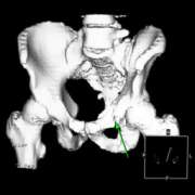
Modern scanners have shortened the time taken to acquire the image and improved the resolution of the axial cuts. Advances in the image reconstruction software has allowed the axial cuts to be rendered as 3-D sections or as slices in planes other than axial. These improvements have allowed CT to be used in areas traditionally the domain of magnetic resonance imaging or nuclear medicine.
Talk Topics
- History of CT and scanner development
- Radiation considerations
- Capabilities and uses of CT
- Interpretation of images
- Tissue attenuation levels
- Windowing
- Contrast media
- Head CT
- Infarct and haemorrhage
- Brain death
- Mass lesions
- Chest CT
- Comparison with chest x-ray
- Mediastinum on CT
- Pulmonary embolus
- Mass lesions
- Abdomen CT
- Comparison with abdominal x-ray
- Pancreatitis
- Pelvic inflammation
- Tumour location and staging
- Traumatic bleeding
- Spine fractures
- Limb fractures