Chest
Given the frequency with which radiology is used to investigate chest pathology, the tutorial has been split into two parts. The first covers an approach to reading a chest x-ray, then progresses to the normal chest CT and then discusses a variety of pathological appearances, before moving on to the appearance of specific clinical conditions.
The second part continues the clinical perspective and shows how the pathological appearances come together in conditions such as heart failure and lobar collapse, and ends with a guide to differentiating the causes of masses on the chest film.
This tutorial provides a solid introduction to radiology of the chest and is complemented by the cardiovascular tutorial and chest pain and trauma tutorial.
Chest 1
- Normal Chest Film
- Normal Variations
- CT Normal Chest
- Pneumothorax
- Pleural Effusions
- Airspace Consolidation
- Interstitial Changes
- Silhouette Diagram
- Lobar Consolidations
- Bronchopneumonia
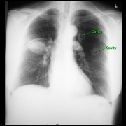
- Abscess
- High-Res CT
- Pulmonary Fibrosis
- High-Res CT and CT Summary
Chest 2
- Heart Failure
- Acute and Chronic
- Collapsed Lung
- Individual Lobes
- Bilateral Collapse
- Plate Atelectasis
- Round Lesions
- Overview of Coin Lesions
- Metastasis
- Wegener’s Granulomatosis
- Arteriovenous Malformation
- Tuberculosis
- Mediastinal Lesions
- Goitre
- Thymoma
- Hodgkin’s Lymphoma
- Bronchial Cyst
- Hiatus Hernia