Magnetic Resonance Imaging
Magnetic resonance imaging uses strong magnetic fields and radiofrequency waves to produce a signal that can be reconstructed into an image slice, much like in computed tomography. It does not use or produce any ionising radiation, as opposed to computed tomography, nuclear medicine and general radiography, although the strong magnetic fields used cause problems for metal implants or foreign bodies, pacemakers and other medical equipment.
A variety of image sequences and timings, combined with contrast agents, makes for detailed imaging of anatomy and pathology. As the technology has matured it has also become possible to reduce the scale of the units, leading to smaller low-resolution magnetic resonance imaging systems suitable for private rooms. This allows magnetic resonance imaging to be used in areas traditionally served by ultrasound.
Talk Topics
- History, principles and advantages of MRI
- Image sequences
- Head MR
- Comparison with CT
- Multiple sclerosis
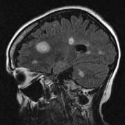
- Intracranial tumours
- Infarct and haemorrhage
- Diffuse axonal injury
- Spine MR
- Spinal cord
- Vertebral fractures
- Infections
- Meningioma
- Musculo-skeletal MR
- Soft-tissue injury
- Joint effusions
- Fractures
- Tumours
- Other uses